BIØLØGLITCHY
The body begins at the cellular level. So does the glitch.
Cells replicate. Most of the time, the copy is accurate. Sometimes — through random mutation, environmental damage, genetic error — the copy is wrong. The cell that results doesn't behave as it should. It divides differently. It survives when it shouldn't. It accumulates. The body doesn't intend this. It happens anyway. What biological research does with staining and fluorescence is illuminate that failure from the inside — applying chemical agents that bind to specific cellular structures and make them glow, so that what is happening can be seen, studied, and if possible, reversed.
BIØLØGLITCHY takes the opposite direction. The image file is intact. It works. Databending breaks it deliberately — passing it through incompatible formats, introducing errors into the data structure, corrupting the pixel information layer by layer. The damage is intentional. And what the damage reveals is the structure that was always there but invisible: the architecture underneath the image, the logic of the file before it became a picture.
Two techniques. Opposite directions. The same result: something invisible becomes visible.
Staining illuminates a system that is breaking down without knowing it. Databending illuminates a system that is working perfectly — by breaking it. One looks at failure to understand it. The other creates failure to see what it exposes. In both cases, what appears is something the original could not show. The cell under fluorescent light. The pixel under format disruption. Structure revealed by damage — biological or digital, involuntary or deliberate.
BIØLØGLITCHY is one more way of looking at the body — the central subject of DISØRDIN∆RY BÆUTY. Not through the portrait, the filter, the beauty standard. Through the microscope. Through images produced by science to study what goes wrong inside the body — tumours, cellular damage, pathology. Images that are, by any conventional definition, images of something ugly. And yet, under fluorescent staining, at cellular scale, they produce something that has its own visual logic. Its own structure. Its own strange beauty. The thorn sense of beauty applied not to the surface of the body but to its interior — to the point where the body is most vulnerable, most itself, most human.
The glitch enters here as method and as parallel. The same images, databended. Two techniques of exposure. Two ways of making visible what the eye cannot reach.
DØM B∆RR∆ — BIØLØGLITCHY — Pluripotent Cells, 2020
Works
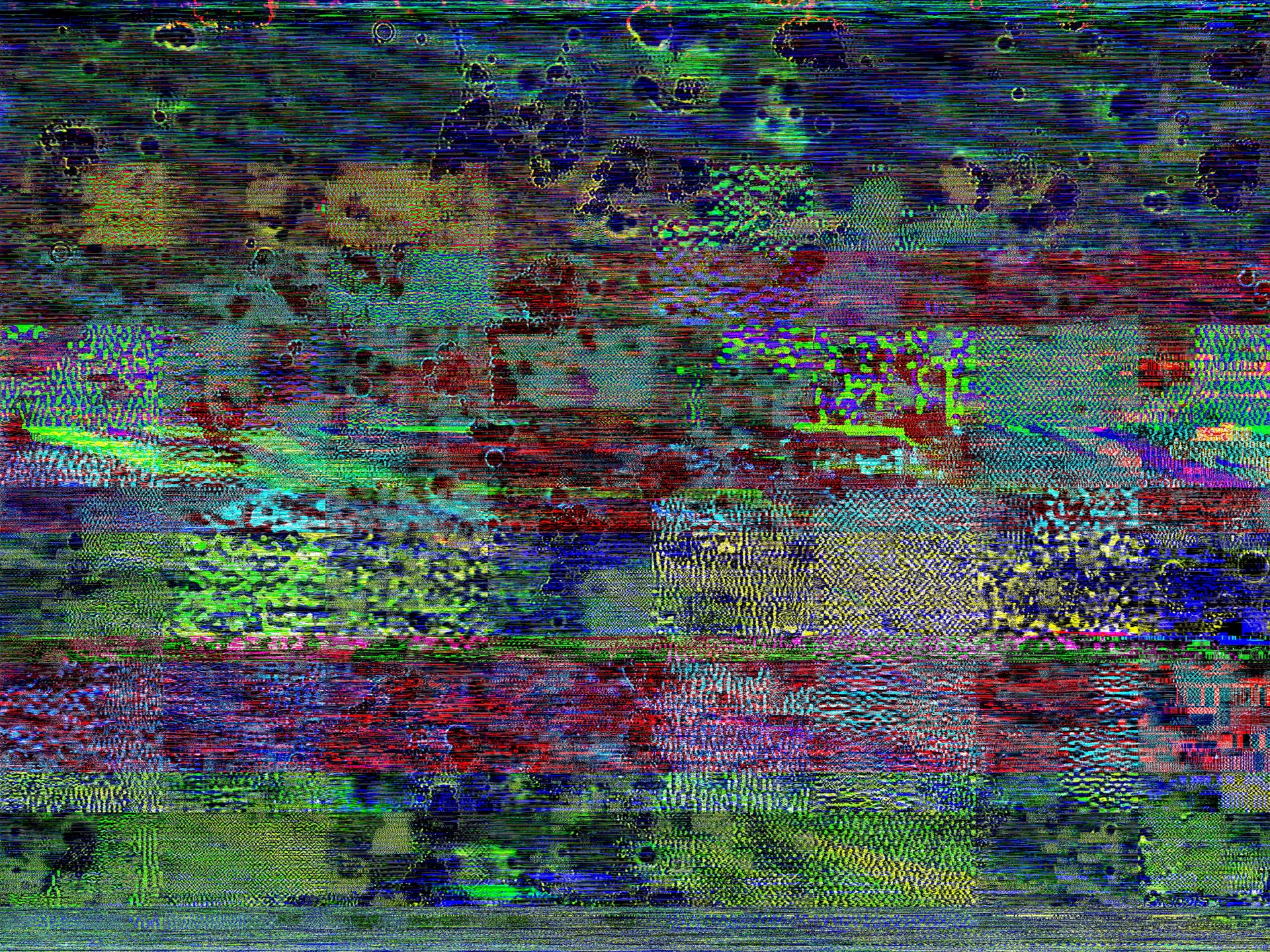
DØM B∆RR∆ — BIØLØGLITCHY — Adipocytes, 2017
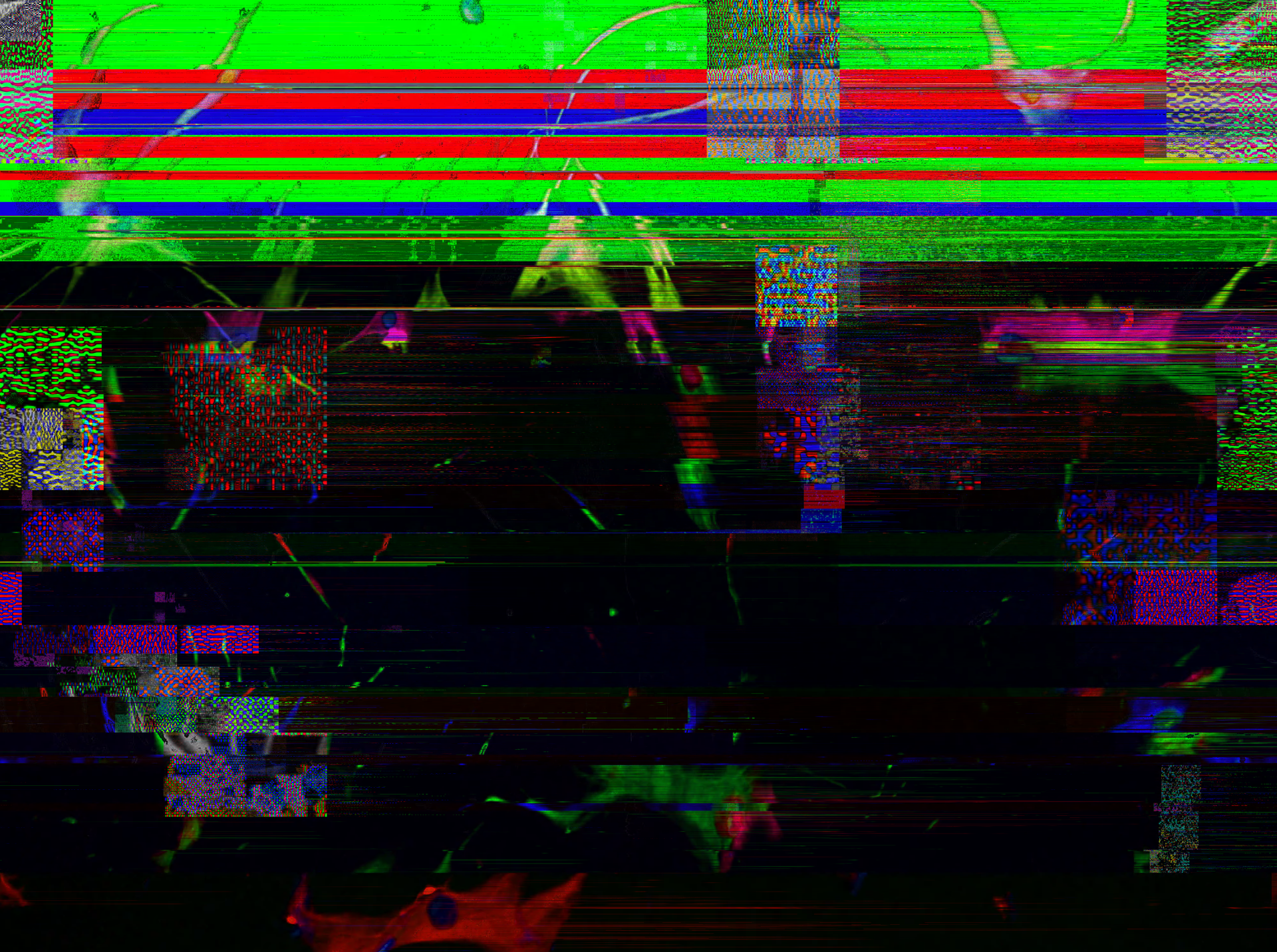
DØM B∆RR∆ — BIØLØGLITCHY — Cells 1, 2018
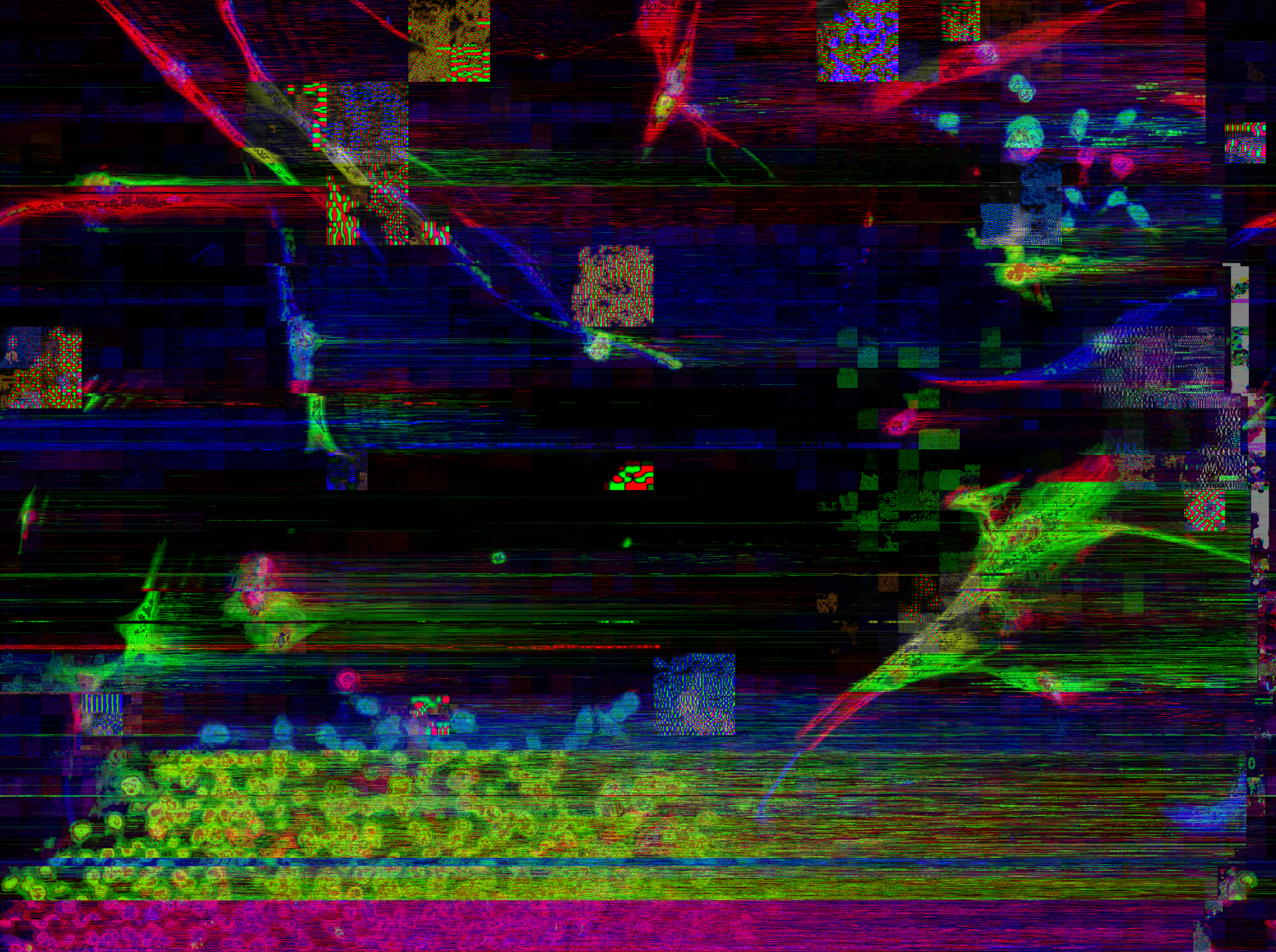
DØM B∆RR∆ — BIØLØGLITCHY — Cells 2, 2018
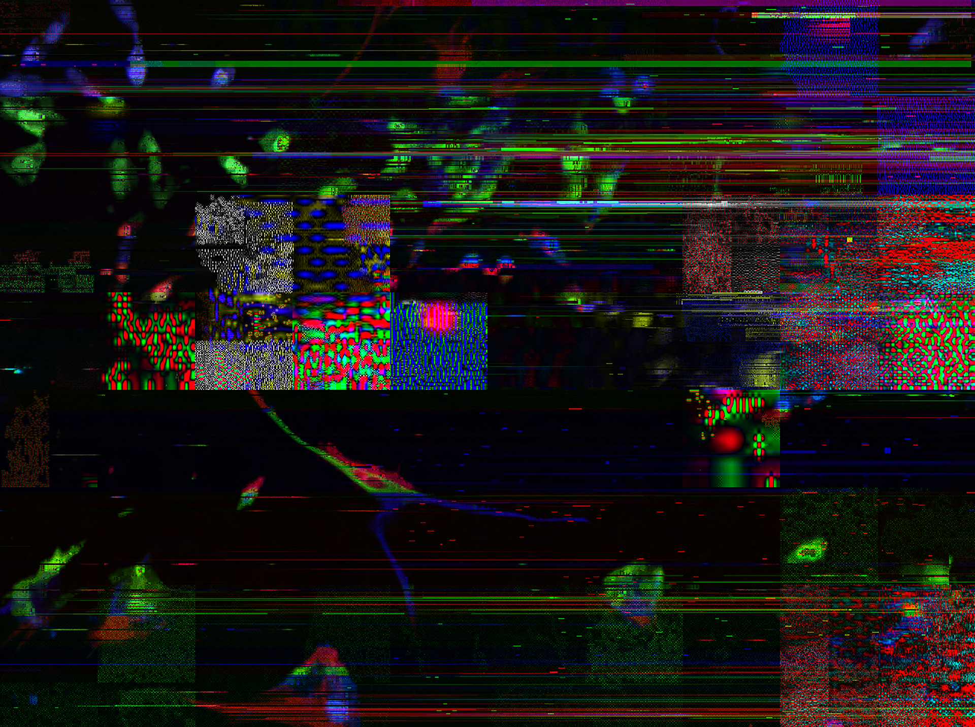
DØM B∆RR∆ — BIØLØGLITCHY — Cells 3, 2018
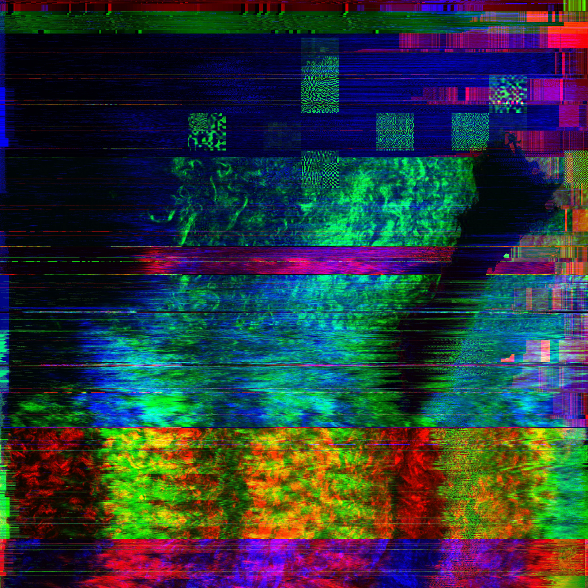
DØM B∆RR∆ — BIØLØGLITCHY — Lungs 1, 2017
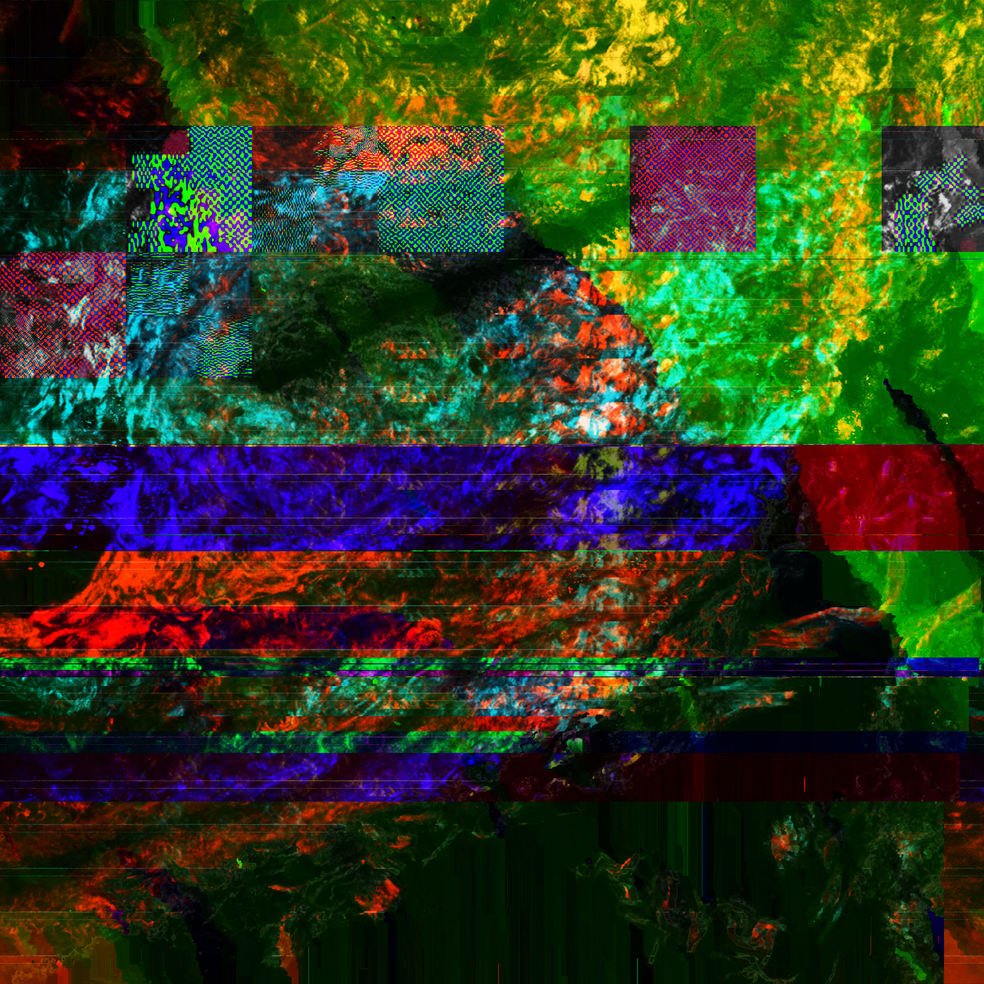
DØM B∆RR∆ — BIØLØGLITCHY — Lungs 2, 2017
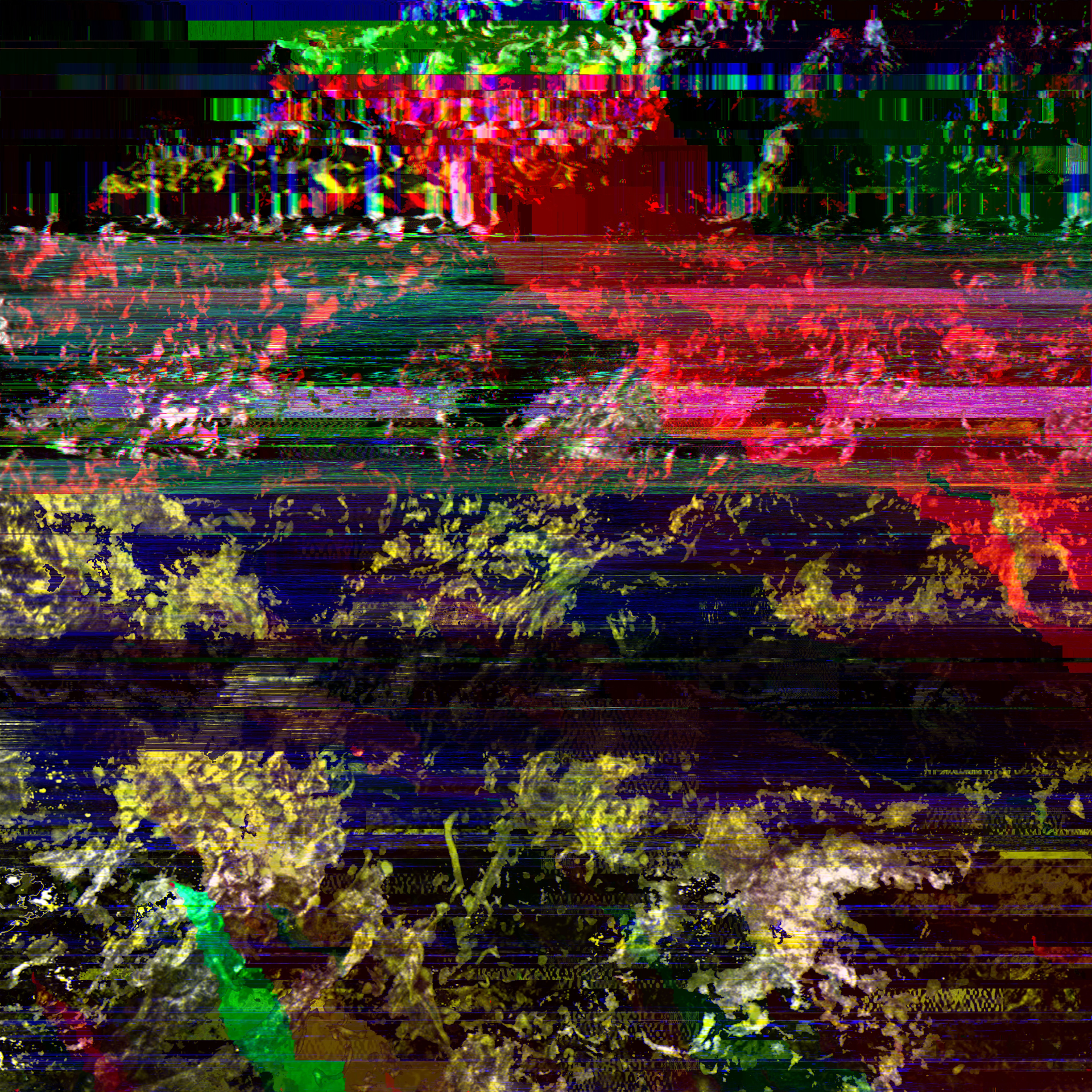
DØM B∆RR∆ — BIØLØGLITCHY — Lungs 3, 2017
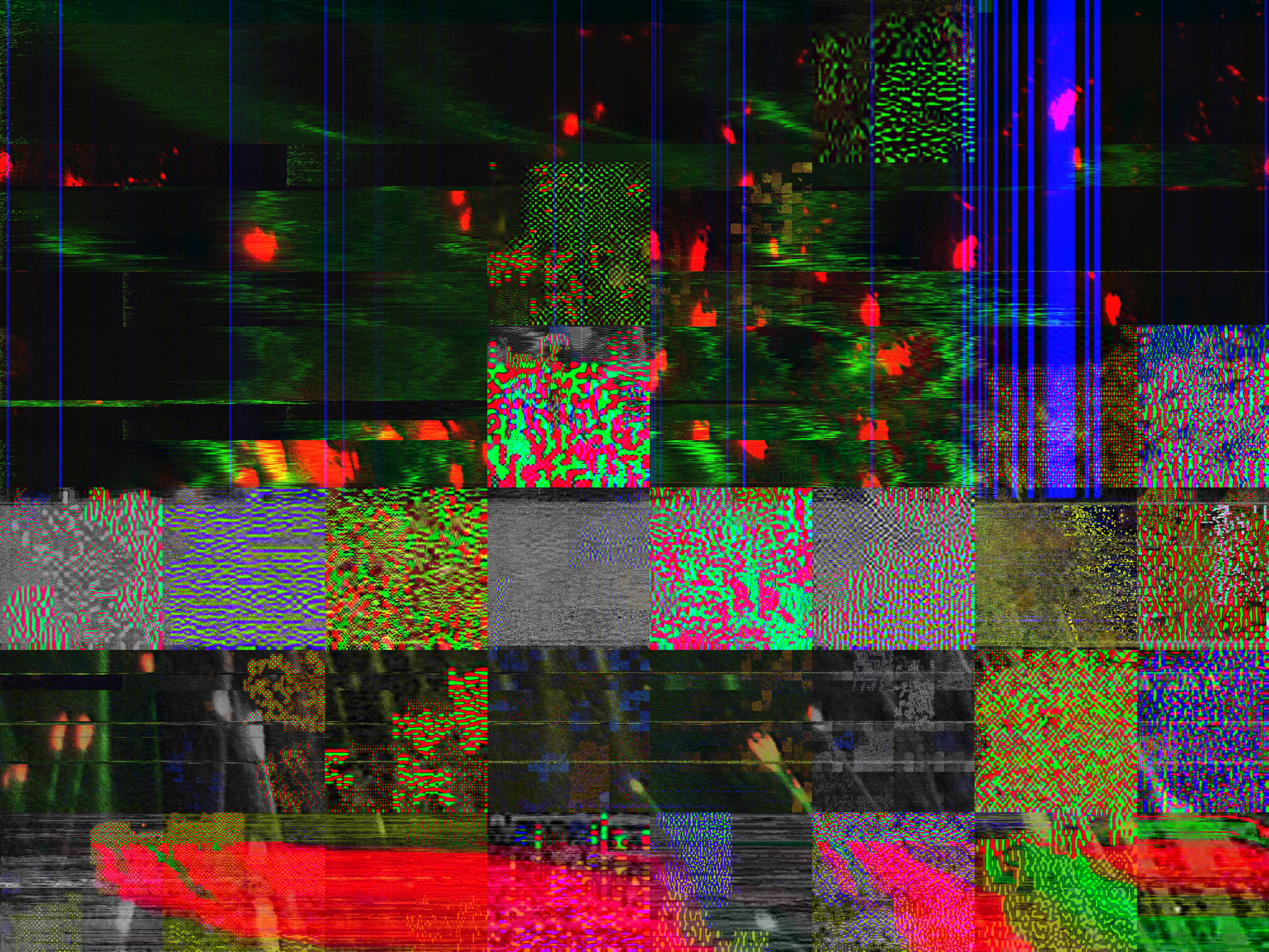
DØM B∆RR∆ — BIØLØGLITCHY — Murine Cells, 2017
On the method
Source: scientific microscopy images of cellular damage and pathology, made visible through fluorescent staining. Process: databending, passing the files through incompatible formats and corrupting the pixel information layer by layer. Two techniques of exposure: one biological, one digital.